Login or Register for free to create your own customized page of blog posts from your favorite blogs. You can also add blogs by clicking the "Add to MyJacketFlap" links next to the blog name in each post.
Blog Posts by Tag
In the past 7 days
Blog Posts by Date
Click days in this calendar to see posts by day or month
Viewing: Blog Posts Tagged with: radiology, Most Recent at Top [Help]
Results 1 - 7 of 7
How to use this Page
You are viewing the most recent posts tagged with the words: radiology in the JacketFlap blog reader. What is a tag? Think of a tag as a keyword or category label. Tags can both help you find posts on JacketFlap.com as well as provide an easy way for you to "remember" and classify posts for later recall. Try adding a tag yourself by clicking "Add a tag" below a post's header. Scroll down through the list of Recent Posts in the left column and click on a post title that sounds interesting. You can view all posts from a specific blog by clicking the Blog name in the right column, or you can click a 'More Posts from this Blog' link in any individual post.
In the wake of the development of advanced neonatal intensive medical care, more and more children born very preterm manage to beat the previously tough odds and survive the perils of infections and respiratory distress that are some of the common problems in the group. While this is one of the success stories of modern medicine, long-term follow-up of premature-born pediatric cohorts show that the obstacles don’t cease with the need of intensive medical care.
The centenary of the Great War has led to a renewed interest in military matters, and throughout history, war has often been the setting for medical innovation with major advances in the treatment of burns, trauma, and sepsis emanating from medical experience in the battlefield. X-rays, which were discovered in 1895 by Roentgen, soon found a role in military conflict. The first use of X-rays in a military setting was during the Italo-Abyssinian war in 1896.
Tomorrow, 8 November, will mark the third anniversary of the now established International Day of Radiology, an event organised by the European Society of Radiology and Radiological Society of North America: a day in which health care workers worldwide mark their debt of gratitude to Wilhelm Roentgen’s great discovery of x- rays, and its subsequent applications in the field of medical practice, today known as radiology or medical imaging. On 8 November 1895, Roentgen conducted his seminal experiment, which was to change the world forever and earn Roentgen the first Nobel Prize for Physics. This day now is celebrated by over one hundred learned radiology societies worldwide to promote the importance of this discipline in current medical practice, a discipline which has changed beyond wildest recognition from the early days of the pioneers and radiation martyrs. The day is a celebration of all radiology team members’ contribution to patient care. In the early days of this new discipline practitioners were not confined to members of the medical profession but included any lay interested member of the public. Only with the passage of time did the discipline of radiology become the sole preserve of medical practitioners, with appropriate training and regulation introduced to raise standards.
It is interesting to note that the first multidisciplinary society devoted to the new subject ‘The Roentgen society’ was founded in 1897 in London by David Walsh, F.E. Fenton, and F. Harrison Low. In the summer of that year, Professor Silvanus Thompson, the physicist, Fellow of the Royal Society of London, brilliant lecturer, populiser of science, prolific author, and true Victorian polymath became its inaugural President. It has since metamorphosed into the current British Institute of Radiology.
One of the celebratory themes of this year’s International Day of Radiology is brain imaging. The immediate early application of x-rays was to look for fractures and localise foreign bodies, leading to their application in the military setting. In the early days, x-rays did not allow doctors to directly visualise the brain. Arthur Schueller, the Viennese radiologist who worked closely with G. Holzknecht, became an early pioneer in using x-rays to make neuroradiological diagnosis and help neurosurgeons deal with brain tumours.
Doctor review brain images by Rhoda Baer. Public domain via Wikimedia Commons
Although the brain itself could not be seen by x-rays, secondary signs from tumours often showed, such as erosion of the skull bones. Localisation of tumours was not an exact science and early detection was difficult. The American Walter Dandy, who worked at Johns Hopkins Hospital, pioneered the imaging of the ventricles by introducing air and contrast, this assisted surgeons in localising tumours of the brain by looking for ventricular displacement. It is claimed that the great Pulitzer Prize-winning neurosurgeon Harvey Cushing thought that this technique would take the skill out of making a diagnosis by clinically examining the patient, though by today’s standards, a clinical examination would not be considered a pleasant investigation to have.
In Portugal in the late 1920s the polymath Egaz Moniz pioneered cerebral angiography, enabling doctors to visualise the blood supply to the brain, including tumours; this was a great step forward. Moniz was an author, researcher, was at one time Portuguese Foreign Secretary, and was also awarded the Nobel Prize in 1949 for his medical advances. However, a really great leap in brain imaging occurred in the early 1970s, when CT scanning (invented by the British genius Hounsfield) came of age, enabling doctors to visualise the brain itself, along with Magnetic Resonance Imaging (MRI) in the 1980s, further clarifying the workings of the normal and abnormal brain.
Today diagnostic imaging, including the more sophisticated CT scanners, are available even in less affluent countries, and their applications and uses in patient care continues to multiply. They have replaced some of the earlier, more dangerous and uncomfortable investigations endured by the preceding generations of patients. More affluent nations continue to see an exponential growth in modern radiological investigations; such is our fascination for high technology.
Today we salute the pioneers in radiology whose efforts have left us with safer, more accurate and more patient friendly tests than ever before. To find our more about International Day of Radiology and its activities, visit the website.
Egyptian mummies continue to fascinate us due to the remarkable insights they provide into ancient civilizations. Flinders Petrie, the first UK chair in Egyptology did not have the luxury of X-ray techniques in his era of archaeological analysis in the late nineteenth century. However, twentieth century Egyptologists have benefited from Roentgen’s legacy. Sir Graham Elliott Smith along with Howard Carter did early work on plain x-ray analysis of mummies when they X-rayed the mummy Tuthmosis in 1904. Numerous X-ray analyses were performed using portable X-ray equipment on mummies in the Cairo Museum.
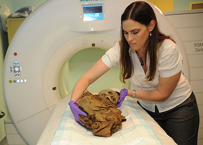
Since then, many studies have been done worldwide, especially with the development of more sophisticated imaging techniques such as CT scanning, invented by Hounsfield in the UK in the 1970s. With this, it became easier to visualize the interiors of mummies, thus revealing their hidden mysteries under their linen wrapped bodies and the elaborate face masks which had perplexed researchers for centuries. Harwood Nash performed one of the earliest head scans of a mummy in Canada in 1977 and Isherwood’s team along with Professor David also performed some of the earliest scannings of mummies in Manchester.
Tori Randall, PhD prepares a 550-year old Peruvian child mummy for a CT scan, by Samantha A. Lewis for the US Navy. Public domain via Wikimedia Commons.
A fascinating new summer exhibition at the British Museum has recently opened, and consists of eight mummies, all from different periods and Egyptian dynasties, that have been studied with the latest dual energy CT scanners. These scanners have 3D volumetric image acquisitions that reveal the internal secrets of these mummies. Mummies of babies and young children are included, as well as adults. There have been some interesting discoveries already, for example, that dental abscesses were prevalent as well as calcified plaques in peripheral arteries, suggesting vascular disease was present in the population who lived over 3,000 years ago. More detailed analysis of bones, including the pelvis, has been made possible by the scanned images, enabling more accurate estimation of the age of death.
Although embalmers took their craft seriously, mistakes did occur, as evidenced by one of the mummy exhibits, which shows Padiamenet’s head detached from the body during the process, the head was subsequently stabilized by metal rods. Padiamenet was a temple doorkeeper who died around 700BC. Mummies had their brains removed with the heart preserved as this was considered the seat of the soul. Internal organs such as the stomach and liver were often removed; bodies were also buried with a range of amulets.
The exhibit provides a fascinating introduction to mummies and early Egyptian life more than 3,000 years ago and includes new insights gleaned from cutting edge twenty first century imaging technology.
Major trauma impacts on the lives of young and old alike. Most of us know or are aware of somebody who has suffered serious injury. In the United Kingdom over five-thousand people die from trauma each year. It is the most common cause of death in people under forty. Many of the fifteen-thousand people who survive major trauma suffer life-changing injuries and some will never fully recover and require life-long care. Globally it is estimated that injuries are responsible for sixteen-thousand deaths per day together with a large burden of people left with permanent disability. These sombre statistics are driving a revolution in trauma care.
A key aspect of the changes in trauma management in the United Kingdom and around the world is the organisation of networks to provide trauma care. People who have been seriously hurt, for example in a road traffic accident, may have suffered a head injury, injuries to the heart and lungs, abdominal trauma, broken limbs, and serious loss of skin and muscle. The care of these injuries may require specialist surgery including neurosurgery, cardiothoracic surgery, general (abdominal and pelvic) surgery, orthopaedic surgery, and plastic surgery. These must be supported by high quality anaesthetic, intensive care, radiological services and laboratory services. Few hospitals are able to provide all of the services in one location. It therefore makes sense for the most seriously injured patients to be transported not to the nearest hospital but to the hospital best equipped to provide the care that they need. Many trauma services around the world now operate on this principle and from 2010 these arrangements have been established in England. Hospitals are designated to one of three tiers: major trauma centres, trauma units, and local emergency hospitals. The most seriously injured patients are triaged to bypass trauma units and local emergency hospitals and are transported directly to major trauma centres. While this is a new system and some major trauma centres in England have only “gone live” in the past two years, it has already had an impact on trauma outcomes, with monitoring by the Trauma Audit and Research Network (TARN) indicating a 19% improvement in survival after major trauma in England.
Not only have there been advances in the organisation of trauma services, but there have also been advances in the immediate clinical management of trauma. In many cases it is appropriate to undertake “early definitive surgery/early total care” – that is, definitive repair of long bone fractures within twenty-four hours of injury. However, patients who have suffered major trauma often have severe physiological and biochemical derangements by the time they arrive at hospital. The concepts of damage control surgery and damage control resuscitation have emerged for the management of these patients. In this approach resuscitation and surgery are directed towards stopping haemorrhage, performing essential life-saving surgery, and stabilising and correcting the patient’s physiological state. This may require periods of surgery followed by intervals for the administration of blood and clotting factors and time for physiological recovery before further surgery is undertaken. The decision as to whether to undertake early definitive care or to institute a damage control strategy can be complex and is made by senior clinicians working together to formulate an overview of the state of the patient.
Modern radiology and clinical imaging has helped to revolutionise modern trauma management. There is increasing evidence to suggest that early CT scanning may improve outcome in the most unstable patients by identifying life-threatening injuries and directing treatment. When a source of bleeding is identified it may be treated surgically, but in many cases interventional radiology with the placement of glue or metal coils into blood vessels to stop the bleeding offers an alternative and less invasive solution.
The evolution of the trauma team is at the core of modern trauma management. Advances in resuscitation, surgery, and imaging have undoubtedly moved trauma care forward. However, the care of the unstable, seriously injured patient is a major challenge. Transporting someone who is suffering serious bleeding to and from the CT scanner requires excellent teamwork; parallel working so that several tasks are carried out at the same time requires coordination and leadership; making the decision between damage control and definitive surgery requires effective joint decision-making. The emergence of modern trauma care has been matched by the development of the modern trauma team and of specialists dedicated to the care of seriously injured patients. It is to this, above all, that the increasing numbers of survivors from serious trauma owe their lives.
Dr Simon Howell is on the Board of the British Journal of Anaesthesia (BJA) and is the Editor of this year’s Postgraduate Educational Issue: Advances in Trauma Care. This issue contains a series of reviews that give an overview of the revolution in trauma care. The reviews expand on a number of presentations that were given at a two-day meeting on trauma care organised by the Royal College of Anaesthetists in the Spring of 2014. They visit aspects of the trauma patient’s journey from the moment of injury to care in the field, on to triage, and arrival in a trauma centre finally to resuscitation and surgical care.
Founded in 1923, one year after the first anaesthetic journal was published by the International Anaesthesia Research Society, the British Journal of Anaesthesia remains the oldest and largest independent journal of anaesthesia. It became the Journal of The College of Anaesthetists in 1990. The College was granted a Royal Charter in 1992. Since April 2013, the BJA has also been the official Journal of the College of Anaesthetists of Ireland and members of both colleges now have online and print access. Although there are links between BJA and both colleges, the Journal retains editorial independence.
In May this year, the American Osler Society held a joint meeting with the London Osler Society and the Japanese Osler Society in Oxford at the Randolph Hotel. The Societies exist to perpetuate the memory of arguably one the most influential physicians of the early twentieth century, and to discuss topics related to Sir William Osler’s interests. It is fitting that this meeting was held in Oxford, where Osler spent his time as the Regius Professor of Medicine having transferred from another great seat of medical learning at Johns Hopkins Medical School in the United States.
Osler was interested in medical education (he produced his classic textbook, which ran to several editions) and set about trying to improve the education of future doctors. Osler’s other great legacy was his combination of superb clinical skills honed by experience not only on the wards but also in the laboratories, and his great interest in the humanities. Osler always tried to combine these two approaches in his work, and much of his writings and aphorisms are as relevant today as when they were first written. Medical students could read Aequanimitas today more than a century after it was written, and would profit from much of the advice to students within this volume of essays and addresses.
Osler had a great interest in the History of Medicine and helped found the history section of the Royal Society of Medicine in London. This scientific section has continued to flourish for over a century. He believed physicians should be well rounded and well read, and that medicine was a calling of both art and science. Although Osler was not against the idea of specialisation in medicine, he was a superb generalist and could manage both adult and child patients. He believed that doctors owed it to themselves to be well versed in the range of disease and illness afflicting mankind. His early interest in comparative pathology during his time at the Montreal Veterinary College prepared him well when dealing with infectious diseases which in the pre-antibiotic area were the scourge of the day, as compared with today in the West where degenerative diseases, cancers and diseases of longevity have overtaken infections as a major killer in the Western world.
The centenary of the Great War is 2014; it was Osler who started a campaign for the compulsory vaccination of soldiers for typhoid, publishing letters in the Times and The British Medical Journal on this topic. That year his literary output also included his Incunabula Medica, a study of 214 of the earliest printed medical books from 1467-1480. Although finished, it was not published until 1923, four years after his death.
Throughout the late twentieth century medicine has continued to super-specialize at an alarming pace throughout the world, driven by the rapid advances in medical diagnosis and treatment. X-rays were only invented in 1895, and the early part of the twentieth century began to see the introduction of chest x-rays into clinical practice. This was still a world away from CT scans, ultrasounds, and MRI scans, which are now de rigueur in the management of patients. Yet in spite of all this progress, disaffection with the medical profession seems rife. Could it be that the general physicians are going to make a comeback? Perhaps a more humanitarian approach to the patient is what is required again, maybe combined with the inexorable technical progress which will undoubtedly continue in the future. Osler would have been amused to see how the wheel of medical fashion has turned full circle.
Arpan K Banerjee qualified in medicine from St Thomas’s Hospital Medical School in London, UK and trained in Radiology at Westminster Hospital and Guys and St Thomas’s Hospital. In 2012 he was appointed Chairman of the British Society for the History of Radiology of which he is a founder member and council member. In 2011 he was appointed to the scientific programme committee of the Royal College Of Radiologists, London. He is the author/co-author of six books including the recent The History of Radiology.
Subscribe to the OUPblog via email or RSS.
Subscribe to only science and medicine articles on the OUPblog via email or RSS.
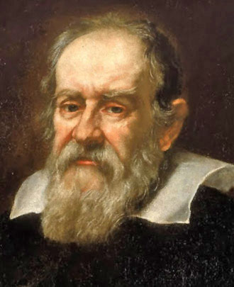
Recently I had the good fortune to see an excellent production of Bertolt Brecht’s play The Life of Galileo at the Birmingham Repertory Theatre. Brecht has a tenuous connection with the medical profession; he registered in 1917 to attend a medical course in Munich and found himself drafted into the army, serving in a military VD clinic for a short while before the end of the war. Brecht’s main interest, however, was drama (in 1918 he wrote his first play Baal) and it was in this field that he made his lasting contribution.
Galileo Galilei
Galileo was persecuted by the Church and the established authorities for his scientific research. His major crime was using his telescope to confirm the Copernican model of the Sun being at the centre of the solar system with the earth revolving around it. This challenged the cultural consensus and the leaders of the day were not prepared to listen to scientific evidence which challenged old dogmas. Galileo was interrogated in the Vatican, tortured, and forced to retract his theories.
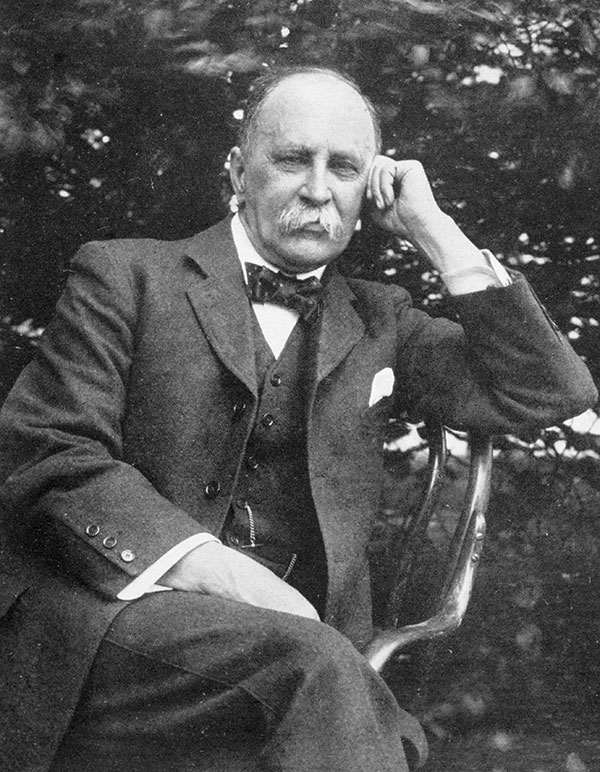
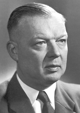
The medical profession has also seen more than its fair share of persecution. I will illustrate with two examples in the relatively new speciality of radiology. The people concerned were not radiologists as such but were conducting pioneering research in imaging. Wilhelm Rötgen, the German physicist who first discovered x-rays in 1895, did himself meet relatively few obstacles regarding the dissemination of his thoughts and findings. But Werner Forssmann, a physician from the small town of Eberswalde in Germany, was not so lucky. In 1929, it is claimed, Forsmann performed the procedure of catheterisation of the heart upon himself and incurred the wrath of his boss as a result. He was sacked and had to switch from a career in cardiology to urology.
Forssmann was to have the last laugh a quarter century later when he shared the 1956 Nobel Prize for his contribution to cardiac catheterisation. This is now a commonplace procedure worldwide.
Werner Forssmann
The next case concerns the plight of Moses Swick, an American urology intern who went to Germany in 1928 to work with Professor Lichtenberg in Berlin. Swick performed scientific studies of a new intravenous contrast agent which would enable visualisation of the renal tract. He and Professor Lichtenberg fell out about who should be given the accolade for the discovery; Lichtenberg stole the limelight and was invited to talk about intravenous urography at the American Urological Association Scientific meeting. For 35 years Swick worked as an urologist in New York until in 1966 it was realised that he had been the victim of injustice and his role in the discovery was belatedly recognised.
These stories are examples where justice prevailed in the end. There are several others which did not and still do not realise fair outcomes.
Arpan K Banerjee qualified in medicine from St Thomas’s Hospital Medical School in London, UK and trained in Radiology at Westminster Hospital and Guys and St Thomas’s Hospital. In 2012 he was appointed Chairman of the British Society for the History of Radiology of which he is a founder member and council member. In 2011 he was appointed to the scientific programme committee of the Royal College Of Radiologists, London. He is the author/co-author of six books including the recent The History of Radiology. Read his previous blog posts.
Subscribe to the OUPblog via email or RSS.
Subscribe to only psychology articles on the OUPblog via email or RSS.
Image credits: (1) Galileo, public domain via Wikimedia Commons. (2) Wilhelm Röntgen, by NFejza, CC-BY-SA-3.0 via Wikimedia Commons. (3) Werner Forssmann nobel, public domain via Wikimedia Commons.


