new posts in all blogs
Viewing: Blog Posts Tagged with: neuroscience, Most Recent at Top [Help]
Results 26 - 35 of 35
How to use this Page
You are viewing the most recent posts tagged with the words: neuroscience in the JacketFlap blog reader. What is a tag? Think of a tag as a keyword or category label. Tags can both help you find posts on JacketFlap.com as well as provide an easy way for you to "remember" and classify posts for later recall. Try adding a tag yourself by clicking "Add a tag" below a post's header. Scroll down through the list of Recent Posts in the left column and click on a post title that sounds interesting. You can view all posts from a specific blog by clicking the Blog name in the right column, or you can click a 'More Posts from this Blog' link in any individual post.

By: Julia Callaway,
on 9/3/2014
Blog:
OUPblog
(
Login to Add to MyJacketFlap)
JacketFlap tags:
Journals,
brain,
neuroscience,
traumatic brain injury,
*Featured,
oxford journals,
Science & Medicine,
Health & Medicine,
Psychology & Neuroscience,
TBI,
concussion,
Louis De Beaumont,
Sébastien Tremblay,
sports injuries,
Add a tag
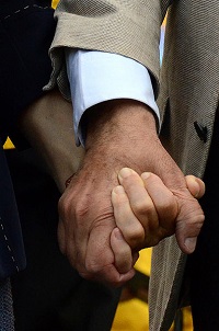
The consequences of traumatic brain injury (TBI) are sizable in both human and economic terms. In the USA alone, about 1.7 million new injuries happen annually, making TBI the leading cause of death and disability in people younger than 35 years of age. Survivors usually exhibit lifelong disabilities involving both motor and cognitive domains, leading to an estimated annual cost of $76.5 billion in direct medical services and loss of productivity in the USA. This issue has received even more intense scrutiny in the popular media with respect to sports-related concussions where there is a proposed link between having suffered multiple injuries, regardless of severity, with later neurodegeneration. At present, there is a dearth of evidence to either support or undermine the role of sports concussions in the later development of neurodegenerative processes, much less the influence of those brain injuries on the normal aging process.
As most people agree that no two concussions are alike, they all share at least one feature in common; they all involve the near instant transfer of kinetic energy to the brain. The brain absorbs kinetic energy as a result of acceleration forces, while deceleration forces cause it to release kinetic energy when colliding with the skull. Coup contrecoup injury is one of the most ancient and best supported biomechanical models of traumatic brain injury induction. Acceleration/deceleration forces can either be transferred to the brain in a straight line passing through the head’s centre of gravity or in a tangential line and arc around its centre of gravity. Shearing and stretching of axons are common manifestations of inertial forces applied to the brain and this type of damage is commonly referred to as traumatic axonal injury. Although robustly demonstrated in both animal and post-mortem models of TBI, neuroimaging techniques limitations, however, have long prevented us from accurately tracking projecting axonal assemblies, also called white matter fibers, in living humans. The recent emergence of a magnetic resonance imaging (MRI)-based tool called Diffusion Tensor Imaging (DTI) can reveal abnormalities in white matter fibers with increasing sensitivity. DTI has quickly gained in popularity among TBI researchers who have long sought to characterize the neurofunctional repercussions of traumatic axonal injury in living humans. One particularly appealing clinical application of DTI is with athletes who have sustained sports concussion in whom conventional MRI assessments typically turn out negative despite the persistence of long-lasting, cumulative neurofunctional symptoms. First applied to young concussed athletes, a follow-up DTI study conducted in our laboratory revealed subtle white matter tracts anomalies detected in the first few days after the injury and again 6 months later. Interestingly, these young concussed athletes were all asymptomatic at follow-up and performance on concussion-sensitive neuropsychological tests had returned to normal.
In parallel, our group became increasingly interested in the characterization of the remote neurofunctional repercussions of concussion sustained decades earlier in late adulthood former elite athletes. Quantifiable cognitive (i.e. memory and attention) and motor function alterations were found on age-sensitive clinical tests, a finding that significantly contrasts with the full recovery typically found within a few days post-concussion in young, active athletes on equivalent neurofunctional measures. This finding was the first of many demonstrations that a remote history of sports concussion synergistically interacts with advancing age to precipitate brain function decline. These neuropsychological tests performance alterations specific to former concussed athletes were soon after found to correlate significantly with markers of structural damage restricted to ventricular enlargement and age-dependent cortical thinning. However, besides the significant interaction of age and a prior history of concussion on cortical thinning, former concussed athletes could not be differentiated from age-matched unconcussed teammates using highly sophisticated measures of grey matter morphometry. White matter integrity disruptions therefore appeared as a likely candidate to explain the observed significant ventricular enlargement found in former concussed athletes. We thus turned to state-of-the-art DTI metrics to conduct the first study of white matter integrity with older but clinically normal retired athletes with a history of sports-related concussions. A particular emphasis was put on bringing together former elite athletes who were free from confounding factors such as clinical comorbidities, drug/alcohol abuse, and genetic predisposition that are too often confusing the long-term effects of concussions on brain health. Our results show that aging with a history of prior sports-related concussions induces a diffuse pattern of white matter anomalies affecting many major inter-hemispheric, intra-hemispheric as well as projection fiber tracts. Of crucial clinical significance with relation to our previous findings on former concussed athletes, we found ventricular enlargement to correlate significantly with widespread alterations of key markers of white matter integrity including not only peri-ventricular white matter tracts, but also an extensive network of fronto-parietal connections. Most of all, these white matter integrity losses were found to be associated with altered neurocognitive functions including memory and learning.
Taken together with previous functional and structural characterizations of the remote effects of concussion in otherwise healthy older former athletes, the pattern of white matter alterations, being more pronounced over fronto-parietal brain areas, more closely resemble what has been observed in normal aging. From this interpretation, we suggest that concussion induces a latent microstructural injury that synergistically interacts with the aging process to exert late-life brain decline in both structure and function.
The post The crossroads of sports concussions and aging appeared first on OUPblog.

By: DanP,
on 8/8/2014
Blog:
OUPblog
(
Login to Add to MyJacketFlap)
JacketFlap tags:
Books,
Music,
creativity,
language,
communication,
jackson,
bilingual,
Michael Jackson,
neuroscience,
genius,
IQ,
hernandez,
*Featured,
Psychology & Neuroscience,
Cognitive Neuroscience,
Arturo Hernandez,
bilingual brain,
arturo,
draehernandez,
child prodigy,
psychometric IQ,
psychometric,
lobes,
Add a tag
By Arturo Hernandez
That any person could become an expert in something if they simply spend about 3 hours per day for ten years learning it is an appealing concept. This idea, first championed by Ericsson and brought to prominence by Gladwell, has now taken root in the popular media. It attempts to discuss these differences in terms of the environment. The idea is that practice with the purpose of constantly gathering feedback and improving can lead any person to become an expert. If becoming an expert requires 10,000 hours, does a prodigy need 20,000.
Lets consider, Michael Jackson, as an example of a prodigy. He grew up in a musical family in Gary, Indiana just outside Chicago. His father Joe played in an R&B band. All of his siblings played music in one way or another. Unlike his siblings and father, Jackson did not really play any instruments. However, he would compose songs in his head using his voice. One morning he came in and had written a song which eventually became ‘Beat It’. In the studio, he would sing each of the different parts including the various instruments. Then the producers and artists in the studio would work on putting the song together, following his arrangements.
Work in cognitive neuroscience has begun to shed light on the brain systems involved in creativity as being linked to psychometric IQ. Work by Neubauer and Fink suggests that these two different types of abilities, psychometric IQ and expertise, involve differential activity in the frontal and parietal lobes. They also appear for different types of tasks. In one study, taxi drivers were split into a high and low group depending on their performance on a paper and pencil IQ test. The results showed that both groups did equally well on familiar routes. The differences appeared between groups when they were compared on unfamiliar routes. In this condition, those with high IQs outperformed those with low IQ. So expertise can develop but the flexibility to handle new situations and improvise requires more than just practice.
Reports of Michael Jackson’s IQ are unreliable. However, he is purported to have had over 10,000 books in his reading collection and to have been an avid reader. His interviews reveal a person who was very eloquent and well spoken. And clearly he was able to integrate various different types of strands of music into interesting novel blends. If we were to lay this out across time, we have perhaps the roots of early genius. It is a person who has an unusual amount of exposure in a domain that starts at an early age. This would lead to the ability to play music very well.

Jackson came from a family filled with many successful musicians. Many were successful as recording artists. Perhaps Michael started earlier than his siblings. One conclusion we can draw from this natural experiment is that creative genius requires more than 10,000 hours. In the case of Michael Jackson, he read profusely and had very rich life experiences. He tried to meld these experiences into a blended musical genre that is uniquely his and yet distinctly resonant with known musical styles.
The kind of creativity is not restricted to prodigies like Michael Jackson. Language, our ultimate achievement as a human race, is something that no other animal species on this planet shares with us. The seeds of language exist all over the animal kingdom. There are birds that can use syntax to create elaborate songs. Chinchillas can recognize basic human speech. Higher primates can develop extensive vocabularies and use relatively sophisticated language. But only one species was able to take all of these various pieces and combine them into a much richer whole. Every human is born with the potential to develop much larger frontal lobes which interconnect with attention, motor, and sensory areas of the brain. It is in these enlarged cortical areas that we can see the roots of creative genius. So while 10,000 hours will create efficiency within restricted areas of the brain, only the use of more general purpose brain areas serve to develop true creativity.
Arturo Hernandez is currently Professor of Psychology and Director of the Developmental Cognitive Neuroscience graduate program at the University of Houston. He is the author of
The Bilingual Brain. His major research interest is in the neural underpinnings of bilingual language processing and second language acquisition in children and adults. He has used a variety of neuroimaging methods as well as behavioral techniques to investigate these phenomena which have been published in a number of peer reviewed journal articles. His research is currently funded by a grant from the National Institutes of Child Health and Human Development. You can follow him on Twitter
@DrAEHernandez. Read his
previous blog posts.
Subscribe to the OUPblog via
email or
RSS.
Subscribe to only brain sciences articles on the OUPblog via
email or
RSS.
Image credit: Michael Jackson with the Reagans, by White House Photo Office. Public domain via Wikimedia Commons.
The post Michael Jackson, 10,000 hours, and the roots of creative genius appeared first on OUPblog.

By: Charley,
on 7/31/2014
Blog:
OUPblog
(
Login to Add to MyJacketFlap)
JacketFlap tags:
Books,
Misinformation,
astrology,
neuroscience,
motto,
*Featured,
Science & Medicine,
Health & Medicine,
Neuroscience in education,
sergio della sala,
pseudoscience,
Psychology & Neuroscience,
sala,
Human Cognitive Neuroscience,
public engageent with science,
public science,
Science festivals,
scientific authority,
sergio,
sergio,
via email or,
Add a tag
By Sergio Della Sala
We are besieged by misinformation on all sides. When this misinformation masquerades as science, we call it pseudoscience. The scientific tradition has methods that offer a way to get accurate evidence and decrease the chance of misinformation persisting for long. The application of these rules marks the difference between science and pseudoscience. Perhaps more importantly, accepting these rules allows us to admit what we do not yet know, and avoids the pomposity too often associated with the notion of scientific authority.

We are easy prey for pseudoscience. We are natural believers, especially in things that we would like to be true. This belief may be fostered by trusting web surfing. We come to believe that our children can improve their scholastic performances by gulping up fishy pills or other improbable supplements. We would like to be more intelligent and show off our skills in solving puzzles, have better memory and absorb volumes of material effortlessly, and to flaunt our astuteness and acumen at parties. To reach these goals by long hours of swotting is a daunting prospect, so we jump at the idea of a quick fix and are prepared to pay for it.
Take the simplistic dichotomy between the two brain hemispheres that informs a series of training programmes. Such programmes are based on the popular assumption that our brains have a nerdy left hemisphere, which acts as a rigorous accountant, opposed to a creative, hippie half, the right hemisphere (which usually needs to be awakened).
Newsmakers fuel belief in tall tales by running uncritical stories advertising outlandish methods and ignoring their obvious flaws. So we can blame the journalists: easy target. However, when we scientists engage with the public, do we really do any better? We are now all desperate to engage the public; our institutions push us to branch out and reach out, and we get brownie points if we do so. This activity too often translates into a scientist going to the media saying “I have nothing to say, and I want to say it on TV.” It sometimes seems that it is the engagement itself that is valued, independently of what we actually say.
There is nothing wrong if you are not interested in science, but if you are then nowadays there are plenty of opportunities to indulge your curiosity. Science festivals are springing up in every city. However, the idea that simply discussing science publicly can counter misinformation is naïve. I posit that too often than it would be advisable, scientists themselves promulgate pseudoscientific thinking, so even science festivals may be counterproductive. Engaging with the public should push scientists to show the evidence and praise scientific methods. We should not abuse the position to dominate by authority.
The Royal Society‘s motto ‘Nullius in verba’ is Latin for, roughly, ‘take nobody’s word for it’. We scientists should remember this motto, not only in our labs, but also when disseminating our ideas. Yet we seem to know no better. Kary Mullis, Nobel Laureate in Chemistry, asserted in his autobiography his belief in astrology. But he is a Capricorn. I’m a Libran, and Librans do not believe in astrology.
Sergio Della Sala is Professor of Human Cognitive Neuroscience at the University of Edinburgh, and co-editor of Neuroscience in Education: The Good, The Bad, and The Ugly.
Subscribe to the OUPblog via email or RSS.
Subscribe to only psychology articles on the OUPblog via email or RSS.
Image credit: Cod liver oil capsules. Photo by Adrian Wold. CC BY 2.5 via Wikimedia Commons.
The post Pseudoscience surplus appeared first on OUPblog.

By: KatherineS,
on 4/10/2014
Blog:
OUPblog
(
Login to Add to MyJacketFlap)
JacketFlap tags:
senses,
neuroscience,
Virtual Reality,
touch,
boundaries,
*Featured,
tactile,
Science & Medicine,
Health & Medicine,
Psychology & Neuroscience,
Cognitive Neuroscience,
multisensory,
gallace,
alberto,
mercier,
bicocca,
Books,
relationships,
Technology,
Add a tag
From Facebook’s purchase of Oculus VR Inc. to the latest medical developments, technology is driving new explorations of the perception, reality, and neuroscience. How do we perceive reality through the sense of touch? Alberto Gallace is a researcher in touch and multisensory integration at the University of Milano-Bicocca, Italy, and co-author of In touch with the future: The sense of touch from cognitive neuroscience to virtual reality. We recently spoke to him about touch, personal boundaries, and being human.
Out of all the human senses, touch is the one that is most often unappreciated, and undervalued. When did you first become interested in touch research?
I was in Oxford as a visiting PhD student and working on multisensory integration, in particular on the integration between tactile and visual signals in the brain. Soon I realized that, despite the fact that is a very important sensory modality, there was not much research on touch, and there were not even a lot of instruments to study such sensory modality. I started by working more with engineers and technical workshops then with psychologists and neuroscientists, just because I needed some device to test the sense of touch in a different way as compared to what was done in the past. Touch was mainly studied with reference to haptic object recognition, mostly on visually impaired individuals or in terms of its physiological mechanisms. Many of the most relevant aspects of touch were very little, if not at all, investigated.
 We use touch for walking, talking, eating, nearly everything basically. It also plays a major role on our interpersonal relationships, it affects the release of hormones and it contributes to define the boundary of our self.
To my students I often say, where our touch begins, we are. I wanted to understand more of these topics. I wanted to compare touch with other sensory modalities. In doing that I was convinced that research on touch had to get away from the fingertips or hands and extend to the whole body surface. The more I studied this sense, the more I became interested in it. For every question answered there were many more without responses. I like touch a lot because there are many things that still need to be understood about it, and I am a rather curious person, particularly when it comes to science.
What do you think has been the most important development in touch research in the past 100 years?
I am not sure if it’s the most important development, but what I certainly consider important is the recent study of certain neural fibres specialized in transmitting socially-relevant information via the sense of touch. That is, the C tactile afferents in humans, that are strongly activated by ‘caress like’ stimuli, might play an important role in many of our most pleasant social experiences. However, I should also say that my personal way to think about science is much more ‘future oriented’. That is, I believe that the most important developments in touch research are the ones that we will see in the next years. I am really looking forward to reading (or possibly writing) about them.
Why did you decide to research this topic?
Most of the previously published books on touch — there aren’t many, to be honest — were focused on a single topic. Most of them were based on research on visually-impaired individuals, and the large majority of them were authored books, a collections of chapters written by different people, sometimes with a different view. Charles [Spence, University of Oxford] and myself wanted something different, something more comprehensive, something that could help people to understand that touch is involved in many different and relevant aspects of our life. We envisioned a book where the more neuroscientific aspects of touch were addressed together with a number of more applied topics. We wanted something where people could see touch ‘at work’. We talk about the neural bases of touch, tactile perception, tactile attention, tactile memory, tactile consciousness, but also about the role of touch in technology, marketing, virtual reality, food appreciation, and sexual behaviour. Many of these topics have never been considered in a book on touch before.

Philippe Mercier’s The Sense of Touch
What do you see as being the future of research in this field in the next decade?
I think that research in my field, pushed by technological advances, will grow rapidly in the coming years. One of the fields where I see a lot of potential is certainly related to the reproduction of tactile sensations in virtual reality environments. Virtual reality will likely become an important part of our life, maybe not in the next decade, but certainly in a not so distant future. However, if we want to create believable virtual environments we need to understand more of our sense of touch, and in particular how our brain processes tactile information, how different tactile stimulations can lead to certain emotions and behaviours, and how tactile sensations can be virtually reproduced. Following the idea that ‘where our touch begins, we are’, research will certainly invest a lot of resources in trying to better understand the neurocognitive mechanisms responsible for supporting our sense of ‘body ownership’ (the feeling that the body is our own) and how this sense can be transferred to virtual/artificial counterparts of our self. Here research on touch will certainly play a leading role.
If you weren’t doing touch research, what would you be doing?
I think I’d work as a scientist in a different field, but always as a scientist. I am too curious about how nature works to do something different. Since I was twelve I’ve always had a special interest in astronomy and astrophysics and I can easily picture myself working in that field too. Understanding the secrets of black cosmic matter or studying the mysteries of white brain matter? Not sure which would be better. What I am sure about is that I like my job a lot, and I won’t change it with anything else that is not based that much on creativity and curiosity.
Alberto Gallace is a researcher at Department of Psychology, University of Milano-Bicocca, Italy, and co-author of In touch with the future: The sense of touch from cognitive neuroscience to virtual reality. His research interests include spatial representation, multisensory integration, tactile perception, tactile interfaces, body representation, virtual reality, sensory substitution systems, and neurological rehabilitation of spatial disorders.
Subscribe to the OUPblog via email or RSS.
Subscribe to only brain sciences articles on the OUPblog via email or RSS.
Image credits: (1) Via Catalana Barcelona Plaça Catalunya 37. Photo by Judesba. CC-BY-SA-3.0 via Wikimedia Commons. (2) The Sense of Touch, painting by Philipe Mercier. Public domain via Wikimedia Commons
The post A conversation with Alberto Gallace appeared first on OUPblog.

By: Julie Fergus,
on 4/9/2014
Blog:
OUPblog
(
Login to Add to MyJacketFlap)
JacketFlap tags:
Earth & Life Sciences,
Psychology & Neuroscience,
flatworms,
model organism,
Oné Pagán,
planarian,
planarian pictures,
The First Brain,
triclads,
pagán,
pagán’s,
planarians,
kawakatsu,
Books,
worms,
neuroscience,
regeneration,
*Featured,
Images & Slideshows,
Science & Medicine,
brain,
masaharu,
“planarian”,
Add a tag
The earth is filled with many types of worms, and the term “planarian” can represent a variety of worms within this diverse bunch of organisms. The slideshow below highlights fun facts about planarians from Oné Pagán’s book, The First Brain: The Neuroscience of Planarians, and provides a glimpse of why scientists like Pagán study these fascinating creatures.
-
Planarian Taxonomy
http://blog.oup.com/wp-content/uploads/2014/04/Planarian-One.jpg
In taxonomic terms, planarians belong to a large class of organisms called Vermes, the Latin term for “worm.” Platyhelminthes, or “flatworms,” represent a phylum within the class of Vermes, though the Platyhelminthes are broken down into four additional categories. One of these categories includes the Turbellarians—flatworms that are free-living and non-parasitic. Turbellarians are then arbitrarily distinguished based on their size, creating two further divisions: the microturbellarians (worms that are shorter than 1 mm) and macroturbellarians (worms that are longer than 1 mm). There are two types of macroturbellarians, the triclads and polyclads. Though “planarian” is a general term used to describe many types of flatworms, it is most often used in reference to triclads.
-
Planarian Size
http://blog.oup.com/wp-content/uploads/2014/04/Planarian-Two.jpg
Triclads are small worms. They do not typically exceed one inch in length, but many of these worms are even smaller than that. Though these planarians can usually be seen with the naked eye, they are often studied under a microscope for closer observation. The two distinctive bumps that you can see on both sides of a planarian’s head, even without a microscope, are called auricles. Though they are commonly mistaken for ears, auricles do not pick up sounds in the environment, and instead contain many chemoreceptors that help planarians sense both nourishing and toxic substances in their surrounding environment.
-
Planarian Fossilization
http://blog.oup.com/wp-content/uploads/2014/04/Planarian-Three.jpg
Planarians are an ancient species. But like other invertebrates, it’s hard for planarians to fossilize because their bodies lack hard bones. Planarians’ tendency to autolyze—or dissolve head first—when they die makes the process of fossilization even more difficult. Though the fossil record for these organisms is a bit scarce, scientists have identified a fully intact turbellarian fossil from the Eocene period about 40 million years ago. Scientists have also found what they interpret as fossilized flatworm tracks from the Permian period (300 million years ago).
-
Planarian Regeneration
http://blog.oup.com/wp-content/uploads/2014/04/Planarian-Four.jpg
Planarians possess the incredible capability to regenerate cells that are damaged or removed. Though the scope of regeneration varies from species to species, many planarians are capable of full regeneration. This means that if you chop up a planarian into several pieces—the current record is 279 sections—each piece can regenerate into a full grown worm, assuming that this piece of the worm is placed in an environment with adequate nourishment. Even the isolated tip of a tail from a planarian can develop into a full grown worm that possesses a brain and central nervous system.
-
Planarians as Model Organisms
http://blog.oup.com/wp-content/uploads/2014/04/Planarian-Five.jpg
In the early 20th century, scientists began looking for organisms with which they could test the principles of Mendelian genetics. Most people are aware that fruit flies (Drosophila melanogaster) became a common test subject because of their short lifespans and ability to produce large quantities of offspring in a short period of time. However, planarians also became a great model organism for scientists to use. Though the planarian nervous system is simple, these flatworms display an immense array of complex behaviors—making planarians an ideal candidate with which scientists can study how the brains of more complex organisms, such as humans, function.
-
Planarians in Popular Culture
http://blog.oup.com/wp-content/uploads/2014/04/Planarian-Six.jpg
Planarians are not only test subjects for scientists to study. These are also creatures of popular culture, making appearances in several movies and TV shows. For example, in an episode of Fringe, Dr. Bishop offers Agent Dunham a smoothie with chopped pieces of planarians—a gesture meant to pay homage to the memory transfer experiments conducted by McConnell. Planarians have also appeared in the first movie of the Twilight saga, in addition to episodes of The Big Bang Theory and Dr. Who.
Oné R. Pagán is a Professor of Biology at West Chester University of Pennsylvania, and the author of The First Brain: The Neuroscience of Planarians.
Subscribe to the OUPblog via email or RSS.
Subscribe to only brain sciences articles on the OUPblog via email or RSS.
Images: The first five photos in this slideshow have been used courtesy of Dr. Masaharu Kawakatsu. Photo six is copyrighted (2003) by the National Academy of Sciences, USA and has been used with permission.
The post Pagán’s planarians: the extraordinary world of flatworms appeared first on OUPblog.

By: Kirsty,
on 4/9/2014
Blog:
OUPblog
(
Login to Add to MyJacketFlap)
JacketFlap tags:
Videos,
Journals,
Multimedia,
brain,
neuroscience,
spinal cord,
Christopher Reeve,
Editor's Picks,
*Featured,
oxford journals,
Science & Medicine,
Health & Medicine,
brain research,
brain journal,
claudia angeli,
complete paralysis,
epidural stimulation,
reeve foundation,
spinal paralysis,
voluntary movement after paralysis,
specific training with epidural stimulation at the human locomotion research cen,
s kentucky spinal cord injury research center,
the first four to undergo task,
left to right is andrew meas,
frazier rehab institute,
as part of the university of louisville,
dustin shillcox,
and rob summers,
Add a tag
By Sam Maddox
Scientists, using epidural stimulation over the lumbar spinal cord, have enabled four completely paralyzed men to voluntarily move their legs.
Kent Stephenson is one of the four. This stimulation experiment wasn’t supposed to work for him; he is what clinicians call an AIS A. This is a measure of disability, formally the American Spinal Injury Association Impairment Scale (AIS), that rates impairment from A (no motor or sensory function) to D (ability to walk). Kent, a mid-thoracic paraplegic, has what is considered a “complete” injury. Kent’s doctors told him it was a waste of time to pursue any therapy; per the dogma, A’s don’t get better. Well, the young Texan, who was hurt five years ago on a dirt bike, didn’t get the message. He likes to cite a fortune cookie he got shortly after his injury. It said, “Everything’s impossible until somebody does it.”
Kent had the stimulator implanted. A few days later they turned it on. No one expected it to do anything. Researchers were only looking for a baseline measurement to compare Kent’s function later, after several weeks of intense Locomotor Training (guided weight supported stepping on a treadmill).
Kent tells the story: “The first time they turned the stim on I felt a charge in my back. I was told to try pull my left leg back, something I had tried without success many times before. So I called it out loud, ‘left leg up.’ This time it worked! My leg pulled back toward me. I was in shock; my mom was in the room and was in tears. Words can’t describe the feeling – it was an overwhelming happiness.”
Click here to view the embedded video.
Kent was the second of the four. Rob Summers, three years ago, was the first to pioneer the concept that complete doesn’t mean what it used to; epidural stimulation could make the spinal cord more receptive to nerve signals coming from the senses or the brain. Seven months after he was implanted with a stimulator unit, he initiated voluntary movements of his legs. The other two subjects, Andrew Meas and Dustin Shillcox, also started moving within days of the implant. Summers probably could have initiated movement early on too, but the research team didn’t test for it – they had no reason to believe he could do it.
Here’s lead author of the Brain paper, Claudia Angeli, Ph.D., to explain. She is a senior researcher at the Human Locomotor Research Center at Frazier Rehab Institute, and an assistant professor at the University of Louisville’s Kentucky Spinal Cord Injury Research Center (KSCIRC).
“First, in the Lancet paper [regarding the first stimulation subject] it was just Rob, just one person. Yes, it was proof of concept, yes it went great. But now we are talking about four subjects. That’s four out of four showing functional recovery. What’s more, two of the four are categorized as AIS A – no motor or sensory function below the lesion level, with no chance for any recovery.”
The other two patients are classified AIS B: no motor function below the lesion but with some sensory function.
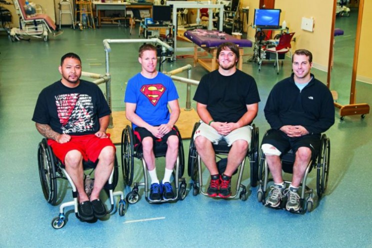
Left to right is Andrew Meas, Dustin Shillcox, Kent Stephenson, and Rob Summers, the first four to undergo task-specific training with epidural stimulation at the Human Locomotion Research Center laboratory, Frazier Rehab Institute, as part of the University of Louisville’s Kentucky Spinal Cord Injury Research Center, Louisville Kentucky.
How does this work? The epidural stimulation supplies a continuous electrical current, at varying frequencies and intensities, to specific locations on the lower part of the spinal cord. A 16-electrode spinal cord stimulator, commonly used to treat pain, is implanted over the spinal cord at T11-L1, a location that corresponds to the complex neural networks that control movement of the hips, knees, ankles and feet.
The leg muscles are not stimulated directly. The epidural stimulation apparently awakens circuitry in the spinal cord. “In simple terms,” says Dr. Angeli, “we are raising the excitability or gain of the spinal cord. Let’s say you have an intent to move. That signal originates in the brain and gets through to the spinal cord but the cord is not aware enough or excited enough to do anything with that intent. When we add the stimulation, the spinal cord networks are made a little more aware, so when the intent comes through, the cord is able to interpret it and movement becomes voluntary.”
The theory behind spinal cord stimulation is that these spinal cord networks are smart: they can remember and they can learn. The current work builds on decades of research. Susan Harkema, Ph.D. (University of Louisville) and V. Reggie Edgerton, Ph.D. (University of California Los Angeles) have led the effort. Dr. Harkema is Principal Investigator for the epidural stimulation projects and Director of the Christopher & Dana Reeve Foundation’s NeuroRecovery Network. Dr. Edgerton, a member of the Reeve Foundation’s International Research Consortium on Spinal Cord Injury, is a basic scientist whose work attempts to understand human locomotion and how the brain and spinal cord adapt and change in response to various interventions, including activity, training and stimulation.
Dr. Harkema says plans are in place to implant eight more patients in the next year. Four will mirror the first group, matched by age, level of injury, time since injury, etc. (Gender, by the way, is not a factor; men with spinal cord injury happen to outnumber women four to one.) Another four patients will be stimulated specifically to control heart rate and blood pressure. Dr. Harkema said one of the first four had issues with low blood pressure. When the stimulator was on, though, the pressure was raised, even without contracting any muscles. They want to assess that sort of autonomic recovery in greater detail.
The research team is aware that epidural stimulation can enhance autonomic function in paralyzed subjects; indeed, the first four subjects report improved temperature control, plus better bowel, bladder, and sexual function. Data is being collected to present that part of the stimulation story in another paper.
Does this mean anyone with a spinal cord injury with an implanted stimulator can move? Not necessarily, says Dr. Harkema. “But what I want people to know about this study is that we need to change our attitude about what a complete injury is, challenge the dogma that in AIS A patients there is no possibility of recovery. The view is that it is not a worthwhile investment to offer even intense rehabilitation to people with complete injuries. They’re not going to recover. But the message now is that there is a tremendous amount available. These individuals have potential for recoveries that will improve their health and quality of life. Now we have a fundamentally new strategy that can dramatically affect recovery of voluntary movement in those with complete paralysis, even years after injury.”
Sam Maddox reports on neuroscience research for the Christopher & Dana Reeve Foundation. He lives in Southern California. The paper about this research, ‘Altering spinal cord excitability enables voluntary movements after chronic complete paralysis in humans‘, appears in Brain: A Journal of Neuroscience.
Brain provides researchers and clinicians with the finest original contributions in neurology. Leading studies in neurological science are balanced with practical clinical articles. Its citation rating is one of the highest for neurology journals, and it consistently publishes papers that become classics in the field.
Subscribe to the OUPblog via email or RSS.
Subscribe to only health and medicine articles on the OUPblog via email or RSS.
Image credit: Video and image both used with permission from the Christopher and Dana Reeve Foundation.
The post Voluntary movement shown in complete paralysis appeared first on OUPblog.

From thoughtful commentary on the history of science to an entertaining blend of science and humor, these blogs have very distinct approaches to science but have one thing in common: a driving curiosity about the world around us.
By: Kirsty,
on 1/17/2013
Blog:
OUPblog
(
Login to Add to MyJacketFlap)
JacketFlap tags:
Medical Mondays,
brain,
neuroscience,
neurology,
Albert Einstein,
cerebral cortex,
*Featured,
Environmental & Life Sciences,
Science & Medicine,
Health & Medicine,
adrianne noe,
brain journal,
dean falk,
einstein's brain,
fred lepore,
frontal lobe,
neuroimaging,
phrenology,
pseudoscience,
einstein’s,
gyri,
cortex,
einstein,
science,
Add a tag
By Dean Falk, Fred Lepore, and Adrianne Noe
Imagine that you return from work to find that a thief has broken into your home. The police arrive and ask if they may dust for finger and palm prints. Which would you do? (A) Refuse permission because palm reading is an antiquated pseudoscience or (B) give permission because forensic dermatoglyphics is sometimes useful for identifying culprits. A similar question may be asked about the photographs of the external surface of Albert Einstein’s brain that recently emerged after being lost to science for over half a century. If asked whether details of Einstein’s cerebral cortex should be identified and interpreted from the photographs, which would you answer? (A) No, because to conduct such a study would engage in the 19th century pseudoscience of phrenology or (B) Yes, because the investigation could produce interesting observations about the cerebral cortex of one of the world’s greatest geniuses and, in light of recent functional neuroimaging studies, might also suggest potentially testable hypotheses regarding Einstein’s brain and those of normal individuals. Lest you think this is a straw man (or Aunt Sally) exercise, more than one pundit has recently invoked the phrenology argument against studying Einstein’s brain. For example, one blogger opines, “I hope no one cares about Einstein’s brain. By this I mean his brain anatomy.” The three of us who were privileged to describe the treasure-trove of recently emerged photographs opted for B.
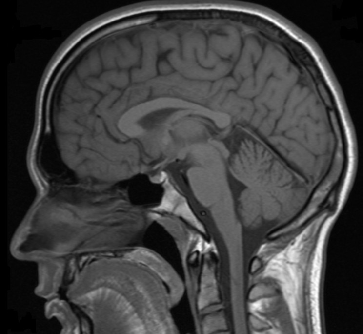
We did so because various data support studying the variation and functional correlates of folds (gyri) on the surface of the human brain and the grooves (sulci) that separate them. For example, David Van Essen hypothesizes that tensions along the connections between cells that course beneath the surface of the brain explains typical patterns of convolutions on its surface. Disruptions in the development of these connections in humans may result in abnormal convolutions that are associated with neurological problems such as autism and schizophrenia. Representations in sensory and motor regions of the cerebral cortex may change later in life as shown by imaging studies of Braille readers, upper limb amputees, and trained musicians, and sometimes these adaptations are correlated with superficial neuroanatomical features such as an enlargement in the right precentral gyrus (called the Omega Sign because of its shape) associated with movement of the left hand in expert string-players. Indeed, Einstein’s right hemisphere has an Omega Sign (labeled K in the following photograph, for knob), which is consistent with the fact that he was a right-handed string-player who took violin lessons between the ages of 6 and 14 years. The aforementioned blogger supports his opposition to studying Einstein’s cerebral cortex with the observation that “assuming causality with correlation” is “a cardinal sin of science”. However, this old saw does not mean that correlated features are necessarily causally unrelated. The functional imaging literature on the cerebral cortices of musicians and controls suggests that Einstein’s Omega Sign and his history as a violinist were probably not an unrelated coincidence.

Superior view of Einstein’s brain, with frontal lobes at the top. The shaded convolution labeled K is the Omega Sign (or knob), which is associated with enlargement of primary motor cortex for the left hand in right-handed experienced string-players.
The investigation of previously unpublished photographs of Einstein’s brain reveals numerous unusual cortical features which suggest hypotheses that others may wish to explore in the histological slides of Einstein’s brain that surfaced along with the photographs. For example, Einstein’s brain has an unusually long midfrontal sulcus that divides the middle frontal region into two distinct gyri (labeled 2 & 3 in the following image), which causes his right frontal lobe to have four rather than the typical three gyri. An extra frontal gyrus is rare, but not unheard of. Einstein’s frontal lobe morphology is interesting because the human frontal polar region expanded differentially during hominin evolution, is involved in higher cognitive functions (including thought experiments), and is associated with complex wiring underneath its surface. These data suggest that the connectivity associated with Einstein’s prefrontal cortex may have been relatively complex, which could potentially be explored by investigating histological slides that were prepared from his brain after it was dissected.

Tracing from photograph of the right side of Einstein’s brain taken with the front of the brain rotated toward viewer. Unusual sulcal patterns are indicated in red; rare gyri are highlighted in yellow.
The microstructural organization in the parts of the cerebral cortex that are involved heavily in speech (Broca’s area and its homologue) were shown to be unique in their patterns of connectivity and lateralization in the genius Emil Krebs, who spoke more than 60 languages. That study, however, did not include information about the gross external neuroanatomy in the relevant regions. Functional neuroimaging technology is making it possible to explore the functional relationships between variations in external cortical morphology, subsurface microstructure including neuronal connectivity, and cognitive abilities. In other words, scientists should now be able to analyze form and function cohesively from the external surface of the cerebral cortex into the depths of the brain. Pseudoscience this is not.
Dr Dean Falk is the Hale G. Smith Professor of Anthropology at Florida State University and a Senior Scholar at the School for Advanced Research in Santa Fe, New Mexico. Dr Fred Lepore is Professor of Neurology and Ophthalmology at Robert Wood Johnson Medical School in Piscataway, New Jersey. Dr Adrianne Noe is Director of the National Museum of Health and Medicine in Silver Spring, Maryland. You can read their paper, ‘The cerebral cortex of Albert Einstein: a description and preliminary analysis of unpublished photographs’ in full and for free. It appears in the journal Brain.
Brain provides researchers and clinicians with the finest original contributions in neurology. Leading studies in neurological science are balanced with practical clinical articles. Its citation rating is one of the highest for neurology journals, and it consistently publishes papers that become classics in the field.
Subscribe to the OUPblog via email or RSS.
Subscribe to only health and medicine articles on the OUPblog via email or RSS.
Image credits: Both images are authors’ own. Do not reproduce without prior permission from the authors.
The post Examining photographs of Einstein’s brain is not phrenology! appeared first on OUPblog.

By: Nicola,
on 1/3/2013
Blog:
OUPblog
(
Login to Add to MyJacketFlap)
JacketFlap tags:
Social Sciences,
Editor's Picks,
*Featured,
and the ugly,
Science & Medicine,
mike anderson,
Neuroscience in education,
sergio della sala,
the bad,
the good,
number nclc,
05276,
teaching,
Education,
Psychology,
neuroscience,
phonics,
neuro,
Add a tag
By Sergio Della Sala & Mike Anderson
In the past ten years, there has been growing interest in applying our knowledge of the human brain to the field of education, including reading, learning, language, and mathematics. Teachers themselves have embraced the neuro revolution enthusiastically. A recent investigation in the US-based journal Mind, Brain, and Education showed that almost 90% of teachers consider knowledge about brain functioning relevant for the planning of education programmes.
This has resulted in the development of a number of new practices in education: some good, some bad, and some just crazy. Too often, people with the clout to make decisions about which practice is potentially profitable in the classroom setting, ignore evidence in favour of gut feelings, the authority of ‘gurus’, or unwarranted convictions. In short, opinions rather than data too often inform implementations in schools. Hence we have had theories suggesting that listening to Mozart can boost intelligence, foot massages can help unruly pupils, fish oil can boost brain power, and even the idea that breathing through your left nostril can enhance creativity! Sadly, it is often scientists themselves who promulgate unsubstantiated procedures.
We shouldn’t ignore the good practices and innovations in education thanks to the developing neuro revolution. A popular example might be the neuroscience data suggesting a strong neural link between fingers and numbers. This is testified by the observation that 6 year old children who are good in recognizing their fingers when touched will later also be better at arithmetical performances. However, more often than not “the good” classroom developments are actually centered around more mainstream cognitive findings. One such finding, named spaced practice, has been replicated many times; it shows that distributing learning over time is more efficient than massing it all together. For example, if students stockpile learning just before an exam, they may do well enough, but if they want to retain the material in the long term, then retrieving it via multiple tests is much better.

Inevitably, we are drawn to discussing “the bad” developments: one of our favourite examples is the use of ineffective coloured lenses to aid reading. This and several other unproven “aids” are potentially damaging the whole idea that knowledge of the mind-brain may contribute to efficacious educational practice. And of course much of current enthusiasm for neuroeducation involves ugly mistranslations of excellent research into an educational arena. Take for instance the misapplication of the well developed theory of reading (the so called dual-route theory) which has been caricatured and wrongly applied in education to justify an ideological stance from teachers preferring a whole-reading (or holistic) approach at the expense of phonics-based teaching. Briefly, the dual-route theory says that single-word reading can be accomplished through a route of letter to sound conversion (phonics) or through a route of direct visual recognition (whole word reading). It does not say that both are equally effective in teaching children to read. Indeed, several studies have demonstrated that phonics is a more effective method; yet the holistic approach to learning to read rages in the classrooms.
The neuro- prefix is very fashionable nowadays, and neuroeducation is just one of the myriad offsprings. Neuroscience offers an invaluable contribution to assess, diagnose, and perhaps manage pathologies, including pathologies of learning in children and adolescents. However, neuroscience as such has so far proved to have little to offer to everyday, normal education. The discipline which has most to offer is instead cognitive psychology, and from this comes some of the “good” that scientists could endow education with. Some of the findings from cognition are solid and counter-intuitive; for example, retrieval practice that, though receiving little support by pedagogists, has proved effective in improving pupils’ learning. This practice is based on the finding that retrieving material through several testing enhances learning of that material more than studying it over and over again.
The psychology of learning could prove efficacious in an educational context. However, science should never be prescriptive; it offers possible windows of knowledge which may or may not be applicable or relevant in specific contexts such as the classroom. There are no ready-made recipes when it comes to mastering the relevance of brain functioning to teaching today. The last thing teachers need is to be superficially trained in neuroscience, but they should certainly watch this space.
Sergio Della Sala is a Clinical Neurologist, Professor of Human Cognitive Neuroscience at the University of Edinburgh, UK. He is co-editor with Mike Anderson of Neuroscience in Education: the good, the bad, and the ugly, and editor of Cortex. His research focuses on the cognitive deficits associated with brain damage.
Mike Anderson is a Professor of Psychology and Director of the Neurocognitive Development Unit at the University of Western Australia. His research focuses on the influence of the developing brain on intellectual functions in children.
Image credit: Photograph of boy studying by Lewis Wickes Hine, ca. 1924, via Library of Congress, Prints & Photographs Division, National Child Labor Committee Collection, [image number nclc.05276].
The post Neuroscience in education appeared first on OUPblog.


Reading level: Ages 9-12
Hardcover: 24 pages
Publisher: Lobster Press (May 25, 2007)
I’ll be the first to admit that I don’t know very much at all about Canadian history, so I was excited when I received a copy of Island of Hope and Sorrow: The Story of Grosse Île the first of Lobster Press’s Canadian Immigration Series.
Organized into different time periods, the book tells us the story of the island, from its first use as a quarantine station for cholera in the early 1800’s to a biological weapons research site during WWII to its opening to the public as a historic site in 1988. Perhaps the most compelling part of the book is the story of Grosse Île’s function as a quarantine site and hospital to care first for European immigrants with cholera and then immigrants struck with typhus as they traveled to Canada on cramped and unsanitary “coffin ships” in attempt to escape the Irish Potato Famine. Located 50 km from Quebec, this was an ideal location to quarantine and treat the sick immigrants and prevent the illnesses from spreading to the colonies. We learn in the book whether or not this was successful and also how, over time, the island advanced.
In addition to the main narrative, the book is loaded with images, journal entries, timelines, posters, and more that give readers a better idea of life on Grosse Île. In addition, a “History Note” sidebar on nearly every page gives tidbits of historical information.
Presenting a human face to immigration, this would make an excellent choice for anyone wanting to learn or teach about immigration, Canadian history, epidemics, and more.
Lookout for the second book in the Canadian Immigration Series: Pier 21: Stories from Near and Far due in April, which I’ll be reviewing soon.


Very cool. I am working on a science blog myself called “The Other Science Blog” combining humor and science.
Thanks Cheri. Lots to explore. The header with coloured flasks is a wonderful photo.
As someone also writing a science blog, it’s nice to see another with humour injected into it, it’s a refreshing change to what I usually read and write
Science blogs are brilliant. If people ever stop being curious then just what is the point?
This planet is the only home we have. Let all citizens work towards saving it for future generations.
Oh wow! Thank you for the recognition. One thing I never anticipated when I started writing was the sheer number of “average” people who were as passionate about science as I am. To see the enquiring mind alive and kicking gives one hope for the human race
This blog is right up my alley because I just wrote the first of several Theoretical Physics essays, although they contain no jokes. There may be some subtle puns, but they contain no-math solutions to the many mysteries about the physical nature of the universe. I explain the current solutions in Modern Physics and offer alternative ideas that to me stay closer to logical thinking in explaining where theory seems to have gone astray.
Thank you for such great effort in sharing science and humor.
Great blogs. Scientific curiosity and inquiry go a long way in unravelling the many mysteries we have about the world around us.
Science blogs are great. There’s nothing we do without a little bit of science