It was midnight and I had just slumped into bed, exhausted after one of my first days on-call as a new intern, and still adjusting to life in a new apartment. As my nagging reflections on the day were just beginning to subside, insistent knocking at my door jolted me back to alertness. Dragging myself out of bed to open the door, I was surprised to see a diminutive elderly lady who appeared quite perturbed.
The post The music next door appeared first on OUPblog.
How would we know if a medieval person had a neurological disorder? If we did know, would it be possible to pinpoint the type of condition? What insight can we gain about the practical impact of disorders on medieval life? Fortunately, a physical record survives that provides a reliable window into the health of medieval people—or, at least, those who were able to write.
The post Scrutinizing the script of the medieval ‘Tremulous Hand of Worcester’ appeared first on OUPblog.
By Sam Maddox
Scientists, using epidural stimulation over the lumbar spinal cord, have enabled four completely paralyzed men to voluntarily move their legs.
Kent Stephenson is one of the four. This stimulation experiment wasn’t supposed to work for him; he is what clinicians call an AIS A. This is a measure of disability, formally the American Spinal Injury Association Impairment Scale (AIS), that rates impairment from A (no motor or sensory function) to D (ability to walk). Kent, a mid-thoracic paraplegic, has what is considered a “complete” injury. Kent’s doctors told him it was a waste of time to pursue any therapy; per the dogma, A’s don’t get better. Well, the young Texan, who was hurt five years ago on a dirt bike, didn’t get the message. He likes to cite a fortune cookie he got shortly after his injury. It said, “Everything’s impossible until somebody does it.”
Kent had the stimulator implanted. A few days later they turned it on. No one expected it to do anything. Researchers were only looking for a baseline measurement to compare Kent’s function later, after several weeks of intense Locomotor Training (guided weight supported stepping on a treadmill).
Kent tells the story: “The first time they turned the stim on I felt a charge in my back. I was told to try pull my left leg back, something I had tried without success many times before. So I called it out loud, ‘left leg up.’ This time it worked! My leg pulled back toward me. I was in shock; my mom was in the room and was in tears. Words can’t describe the feeling – it was an overwhelming happiness.”
Click here to view the embedded video.
Kent was the second of the four. Rob Summers, three years ago, was the first to pioneer the concept that complete doesn’t mean what it used to; epidural stimulation could make the spinal cord more receptive to nerve signals coming from the senses or the brain. Seven months after he was implanted with a stimulator unit, he initiated voluntary movements of his legs. The other two subjects, Andrew Meas and Dustin Shillcox, also started moving within days of the implant. Summers probably could have initiated movement early on too, but the research team didn’t test for it – they had no reason to believe he could do it.
Here’s lead author of the Brain paper, Claudia Angeli, Ph.D., to explain. She is a senior researcher at the Human Locomotor Research Center at Frazier Rehab Institute, and an assistant professor at the University of Louisville’s Kentucky Spinal Cord Injury Research Center (KSCIRC).
“First, in the Lancet paper [regarding the first stimulation subject] it was just Rob, just one person. Yes, it was proof of concept, yes it went great. But now we are talking about four subjects. That’s four out of four showing functional recovery. What’s more, two of the four are categorized as AIS A – no motor or sensory function below the lesion level, with no chance for any recovery.”
The other two patients are classified AIS B: no motor function below the lesion but with some sensory function.
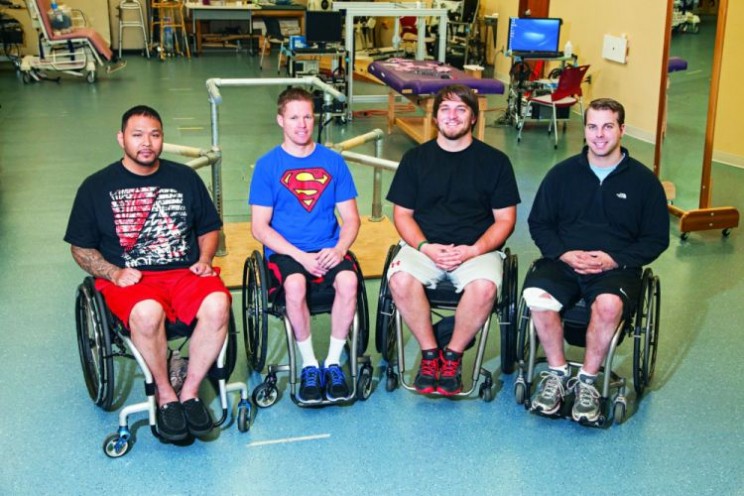
Left to right is Andrew Meas, Dustin Shillcox, Kent Stephenson, and Rob Summers, the first four to undergo task-specific training with epidural stimulation at the Human Locomotion Research Center laboratory, Frazier Rehab Institute, as part of the University of Louisville’s Kentucky Spinal Cord Injury Research Center, Louisville Kentucky.
How does this work? The epidural stimulation supplies a continuous electrical current, at varying frequencies and intensities, to specific locations on the lower part of the spinal cord. A 16-electrode spinal cord stimulator, commonly used to treat pain, is implanted over the spinal cord at T11-L1, a location that corresponds to the complex neural networks that control movement of the hips, knees, ankles and feet.
The leg muscles are not stimulated directly. The epidural stimulation apparently awakens circuitry in the spinal cord. “In simple terms,” says Dr. Angeli, “we are raising the excitability or gain of the spinal cord. Let’s say you have an intent to move. That signal originates in the brain and gets through to the spinal cord but the cord is not aware enough or excited enough to do anything with that intent. When we add the stimulation, the spinal cord networks are made a little more aware, so when the intent comes through, the cord is able to interpret it and movement becomes voluntary.”
The theory behind spinal cord stimulation is that these spinal cord networks are smart: they can remember and they can learn. The current work builds on decades of research. Susan Harkema, Ph.D. (University of Louisville) and V. Reggie Edgerton, Ph.D. (University of California Los Angeles) have led the effort. Dr. Harkema is Principal Investigator for the epidural stimulation projects and Director of the Christopher & Dana Reeve Foundation’s NeuroRecovery Network. Dr. Edgerton, a member of the Reeve Foundation’s International Research Consortium on Spinal Cord Injury, is a basic scientist whose work attempts to understand human locomotion and how the brain and spinal cord adapt and change in response to various interventions, including activity, training and stimulation.
Dr. Harkema says plans are in place to implant eight more patients in the next year. Four will mirror the first group, matched by age, level of injury, time since injury, etc. (Gender, by the way, is not a factor; men with spinal cord injury happen to outnumber women four to one.) Another four patients will be stimulated specifically to control heart rate and blood pressure. Dr. Harkema said one of the first four had issues with low blood pressure. When the stimulator was on, though, the pressure was raised, even without contracting any muscles. They want to assess that sort of autonomic recovery in greater detail.
The research team is aware that epidural stimulation can enhance autonomic function in paralyzed subjects; indeed, the first four subjects report improved temperature control, plus better bowel, bladder, and sexual function. Data is being collected to present that part of the stimulation story in another paper.
Does this mean anyone with a spinal cord injury with an implanted stimulator can move? Not necessarily, says Dr. Harkema. “But what I want people to know about this study is that we need to change our attitude about what a complete injury is, challenge the dogma that in AIS A patients there is no possibility of recovery. The view is that it is not a worthwhile investment to offer even intense rehabilitation to people with complete injuries. They’re not going to recover. But the message now is that there is a tremendous amount available. These individuals have potential for recoveries that will improve their health and quality of life. Now we have a fundamentally new strategy that can dramatically affect recovery of voluntary movement in those with complete paralysis, even years after injury.”
Sam Maddox reports on neuroscience research for the Christopher & Dana Reeve Foundation. He lives in Southern California. The paper about this research, ‘Altering spinal cord excitability enables voluntary movements after chronic complete paralysis in humans‘, appears in Brain: A Journal of Neuroscience.
Brain provides researchers and clinicians with the finest original contributions in neurology. Leading studies in neurological science are balanced with practical clinical articles. Its citation rating is one of the highest for neurology journals, and it consistently publishes papers that become classics in the field.
Subscribe to the OUPblog via email or RSS.
Subscribe to only health and medicine articles on the OUPblog via email or RSS.
Image credit: Video and image both used with permission from the Christopher and Dana Reeve Foundation.
The post Voluntary movement shown in complete paralysis appeared first on OUPblog.

By Dean Falk, Fred Lepore, and Adrianne Noe
Imagine that you return from work to find that a thief has broken into your home. The police arrive and ask if they may dust for finger and palm prints. Which would you do? (A) Refuse permission because palm reading is an antiquated pseudoscience or (B) give permission because forensic dermatoglyphics is sometimes useful for identifying culprits. A similar question may be asked about the photographs of the external surface of Albert Einstein’s brain that recently emerged after being lost to science for over half a century. If asked whether details of Einstein’s cerebral cortex should be identified and interpreted from the photographs, which would you answer? (A) No, because to conduct such a study would engage in the 19th century pseudoscience of phrenology or (B) Yes, because the investigation could produce interesting observations about the cerebral cortex of one of the world’s greatest geniuses and, in light of recent functional neuroimaging studies, might also suggest potentially testable hypotheses regarding Einstein’s brain and those of normal individuals. Lest you think this is a straw man (or Aunt Sally) exercise, more than one pundit has recently invoked the phrenology argument against studying Einstein’s brain. For example, one blogger opines, “I hope no one cares about Einstein’s brain. By this I mean his brain anatomy.” The three of us who were privileged to describe the treasure-trove of recently emerged photographs opted for B.
We did so because various data support studying the variation and functional correlates of folds (gyri) on the surface of the human brain and the grooves (sulci) that separate them. For example, David Van Essen hypothesizes that tensions along the connections between cells that course beneath the surface of the brain explains typical patterns of convolutions on its surface. Disruptions in the development of these connections in humans may result in abnormal convolutions that are associated with neurological problems such as autism and schizophrenia. Representations in sensory and motor regions of the cerebral cortex may change later in life as shown by imaging studies of Braille readers, upper limb amputees, and trained musicians, and sometimes these adaptations are correlated with superficial neuroanatomical features such as an enlargement in the right precentral gyrus (called the Omega Sign because of its shape) associated with movement of the left hand in expert string-players. Indeed, Einstein’s right hemisphere has an Omega Sign (labeled K in the following photograph, for knob), which is consistent with the fact that he was a right-handed string-player who took violin lessons between the ages of 6 and 14 years. The aforementioned blogger supports his opposition to studying Einstein’s cerebral cortex with the observation that “assuming causality with correlation” is “a cardinal sin of science”. However, this old saw does not mean that correlated features are necessarily causally unrelated. The functional imaging literature on the cerebral cortices of musicians and controls suggests that Einstein’s Omega Sign and his history as a violinist were probably not an unrelated coincidence.

Superior view of Einstein’s brain, with frontal lobes at the top. The shaded convolution labeled K is the Omega Sign (or knob), which is associated with enlargement of primary motor cortex for the left hand in right-handed experienced string-players.
The investigation of previously unpublished photographs of Einstein’s brain reveals numerous unusual cortical features which suggest hypotheses that others may wish to explore in the histological slides of Einstein’s brain that surfaced along with the photographs. For example, Einstein’s brain has an unusually long midfrontal sulcus that divides the middle frontal region into two distinct gyri (labeled 2 & 3 in the following image), which causes his right frontal lobe to have four rather than the typical three gyri. An extra frontal gyrus is rare, but not unheard of. Einstein’s frontal lobe morphology is interesting because the human frontal polar region expanded differentially during hominin evolution, is involved in higher cognitive functions (including thought experiments), and is associated with complex wiring underneath its surface. These data suggest that the connectivity associated with Einstein’s prefrontal cortex may have been relatively complex, which could potentially be explored by investigating histological slides that were prepared from his brain after it was dissected.

Tracing from photograph of the right side of Einstein’s brain taken with the front of the brain rotated toward viewer. Unusual sulcal patterns are indicated in red; rare gyri are highlighted in yellow.
The microstructural organization in the parts of the cerebral cortex that are involved heavily in speech (Broca’s area and its homologue) were shown to be unique in their patterns of connectivity and lateralization in the genius Emil Krebs, who spoke more than 60 languages. That study, however, did not include information about the gross external neuroanatomy in the relevant regions. Functional neuroimaging technology is making it possible to explore the functional relationships between variations in external cortical morphology, subsurface microstructure including neuronal connectivity, and cognitive abilities. In other words, scientists should now be able to analyze form and function cohesively from the external surface of the cerebral cortex into the depths of the brain. Pseudoscience this is not.
Dr Dean Falk is the Hale G. Smith Professor of Anthropology at Florida State University and a Senior Scholar at the School for Advanced Research in Santa Fe, New Mexico. Dr Fred Lepore is Professor of Neurology and Ophthalmology at Robert Wood Johnson Medical School in Piscataway, New Jersey. Dr Adrianne Noe is Director of the National Museum of Health and Medicine in Silver Spring, Maryland. You can read their paper, ‘The cerebral cortex of Albert Einstein: a description and preliminary analysis of unpublished photographs’ in full and for free. It appears in the journal Brain.
Brain provides researchers and clinicians with the finest original contributions in neurology. Leading studies in neurological science are balanced with practical clinical articles. Its citation rating is one of the highest for neurology journals, and it consistently publishes papers that become classics in the field.
Subscribe to the OUPblog via email or RSS.
Subscribe to only health and medicine articles on the OUPblog via email or RSS.
Image credits: Both images are authors’ own. Do not reproduce without prior permission from the authors.
The post Examining photographs of Einstein’s brain is not phrenology! appeared first on OUPblog.


